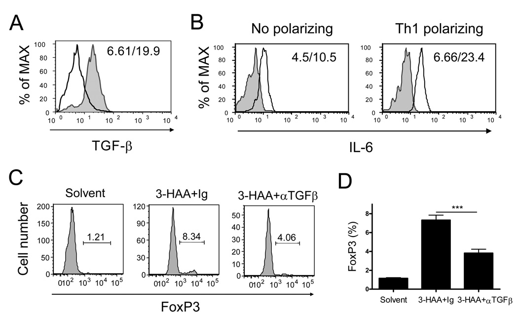
Figure 7. 3-HAA enhances anti-CD3/anti-CD28-induced TGF-β expression and reduces IL-6 production in spleen cells.
A. TGF-β-expression in DCs. Spleen cells from naïve mice were stimulated and primed with either 3-HAA or vehicle control as described in Fig. 5. Cells were harvested from culture on day 2 after stimulation. To determine TGF-β expression, cells were stained with anti-CD11c-FITC, followed by intracellular staining with anti-TGF-β-PerCP/Cy5.5. Data were analyzed by flow cytometry. Data shown are a representative of three experiments. Solid line: vehicle-treated control cells; shaded gray graph: 3-HAA-treated cells. The numbers in each panel represent the mean fluorescence intensity (vehicle/3-HAA). B. IL-6 production in anti-CD3/anti-CD28-activated spleen cells. Spleen cells were stimulated with or without the Th1-polarizing condition, and primed with 3-HAA or vehicle. Supernatants were harvested from culture on day 3 after stimulation. IL-6 levels in culture supernatants were detected by flow cytometry with cytokine bead assay (CBA). Results are shown as graphs reflecting the relative levels of IL-6 in samples. One representative of three experiments is shown. Solid line: vehicle-treated cells; shaded gray graph: 3-HAA-treated cells. The numbers in the panels represent the mean fluorescence intensity (3-HAA/vehicle). C and D. Addition of anti-TGF-b antibody decreased 3-HA-induced FoxP3 expression. Spleen cells were stimulated with anti-CD3/anti-CD28 and primed with 3-HAA (or vehicle) as described in Fig. 6 in the presence of anti-TGF-β antibody (30 µg/ml) or the same amount of Ig control. FoxP3 expression in primed cells was determined as described in Fig. 6. Data are the percentage of FoxP3+ cells in the CD4+CD25+ gate (C) or the mean percentage ± SD of triplicates (D). The results are from a representative of two repeat experiments. ***p < 0.001.

