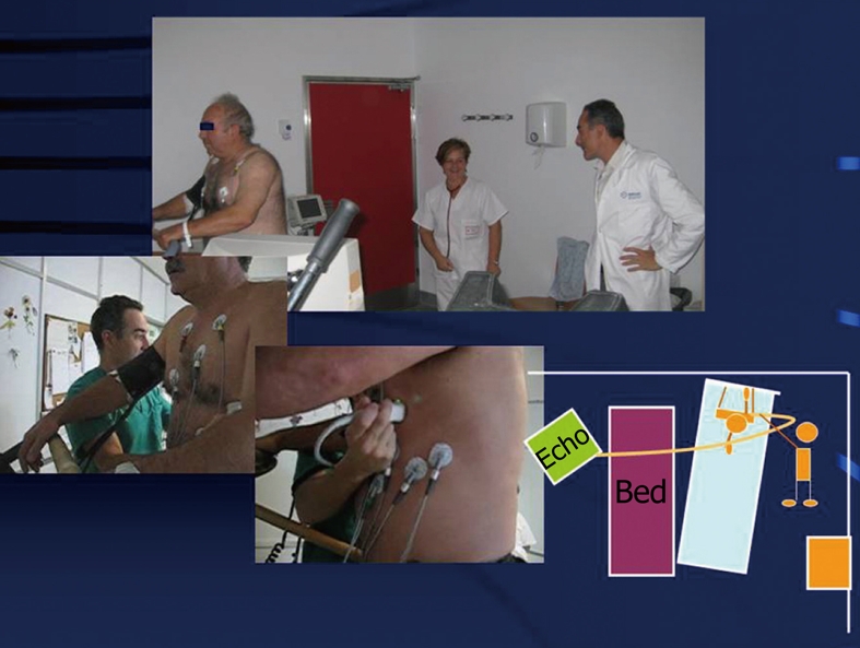
Figure 1.

Exercise echocardiography. When the patient is exhausted or termination criteria appear (symptoms, significative ST changes, decrease or increase in blood pressure, etc.), the observer acquires images by placing the transducer in the cardiac apex, then in the parasternal region. Note the placement of the table, the treadmill and the echocardiography machine for feasible imaging evaluation at peak and post-exercise. The left lateral handlebar of the treadmill has been removed to allow for rapid post-exercise positioning of the patient on the table.
