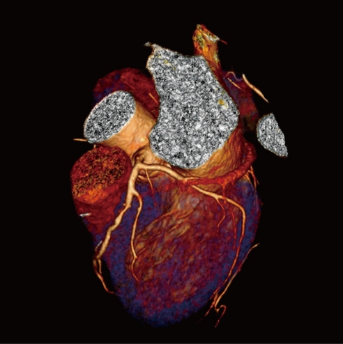
Figure 2.
Three-dimensional volume rendering of the left coronary artery is acquired with dual-source computed tomography angiography in a 47-year-old woman suspected of coronary artery disease. Left anterior descending and left circumflex are clearly demonstrated without any sign of lumen stenosis or calcification.

