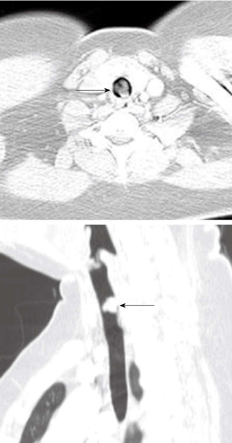
Figure 17.

Axial and sagittal reconstruction of the computed tomography scan images demonstrates a fleshy mass within the tracheal lumen (arrows) in a patient presenting with increasing difficulty of breathing. Histopathology following surgical resection demonstrated a primary sarcoma of the trachea.
