Figure 2.

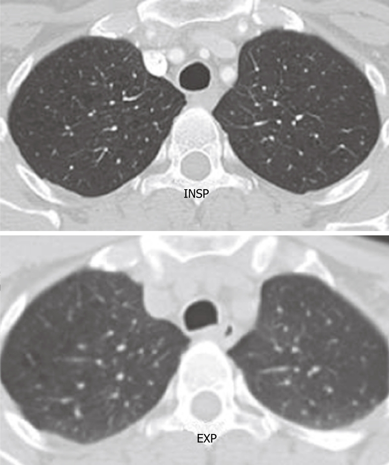
Axial computed tomography image shows the normal rounded configuration of the trachea at the end of inspiration. Note the normal anterior bowing of the posterior membranous wall of the trachea at the end of expiration.

Axial computed tomography image shows the normal rounded configuration of the trachea at the end of inspiration. Note the normal anterior bowing of the posterior membranous wall of the trachea at the end of expiration.