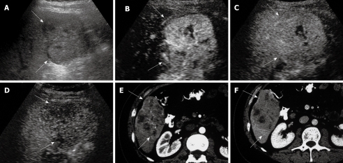
Figure 1.
Hepatocellular carcinoma. A: Gray-scale sonogram shows an isoechogenic nodule (arrows); B: Contrast-enhanced ultrasound (CEUS) scan at arterial phase shows homogeneous hyper-enhancement (arrows); C: CEUS scan at portal phase shows iso-enhancement in comparison with adjacent-liver tissue (arrows); D: CEUS scan at late phase shows hypo-enhancement in comparison with adjacent-liver tissue (arrows); E: Computed tomography (CT) scan shows hyper-attenuation of the nodule (arrows) during the arterial phase; F: CT scan during the portal phase shows hypo-attenuation (arrows).

