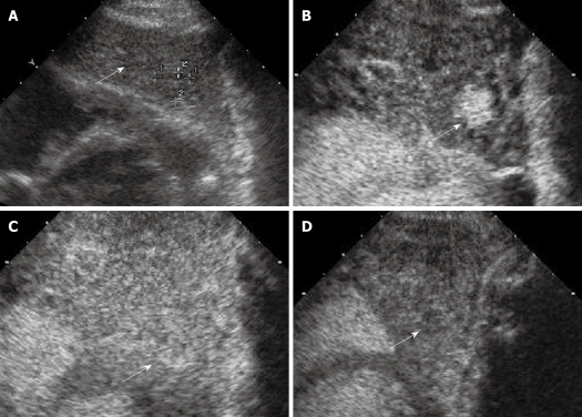
Figure 5.
Biopsy-proven dysplastic nodule. A: A well-defined hypo-echoic lesion in the presence of liver cirrhosis (arrow); B: At CEUS during the arterial phase the lesion shows hyper-enhancement (arrow); C: The lesion appears iso-enhanced with respect to surrounding liver parenchyma in the portal phase(arrow); D: The lesion appears hypo-enhanced with respect to surrounding liver parenchyma in later part of the phase (arrow).

