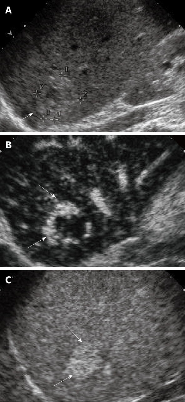
Figure 6.

Hemangioma. A: Gray-scale sonogram shows a hyper-echoic nodule (arrow); B: CEUS scan obtained in arterial phase shows peripheral nodular hyper-enhancement (arrows); C: CEUS scan at late phase shows progressive centripetal enhancement and sustained and complete hyperenhancement (arrows).
