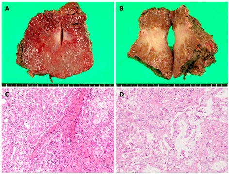
Figure 3.
Gross findings of the resected S3 subsegment. A: The cut surface of the tumor on the ventral side of S3; B: The cut surface of the tumor on the dorsal side of S3. Histopathological findings of the two resected tumors; C: The tumor on the ventral side was pathologically diagnosed as a moderately differentiated hepatocellular carcinoma (HCC) (with nodular, trabecular, and plate-like components); D: The tumor on the dorsal side was pathologically diagnosed as a cholangiocellular carcinoma (CCC) (diffuse type showing a vestigial remnant of the tumor).

