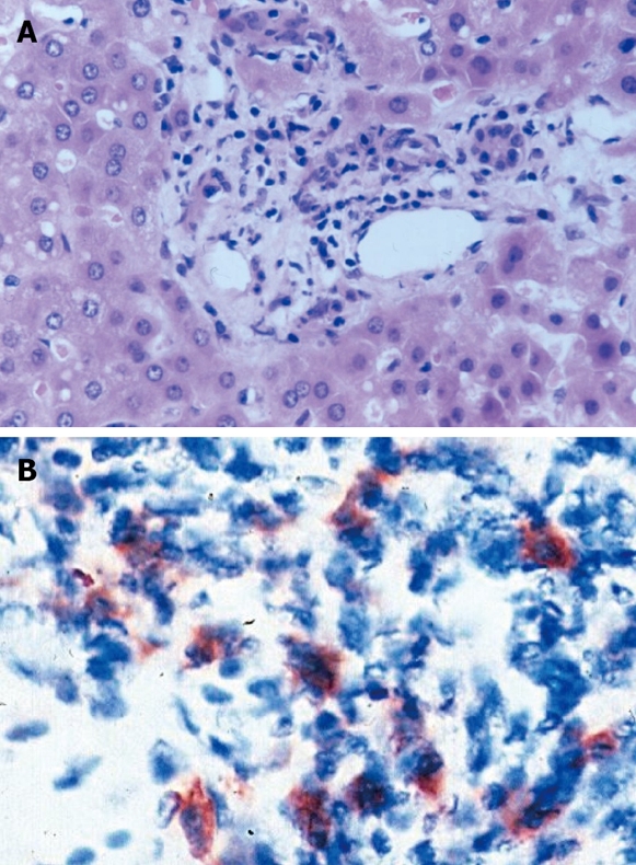
Figure 1.

The histological findings associated with intragraft human herpes virus 6 infection. A: Portal area with mild lymphocyte dominated inflammatory infiltrate (H&E staining, original magnification × 400); B: Human herpes virus 6 positive cells in the portal area demonstrated by immunohistochemistry (original magnification × 1000). From Härmä et al. Transplantation 2006; 81: 367-372 with permission[52].
