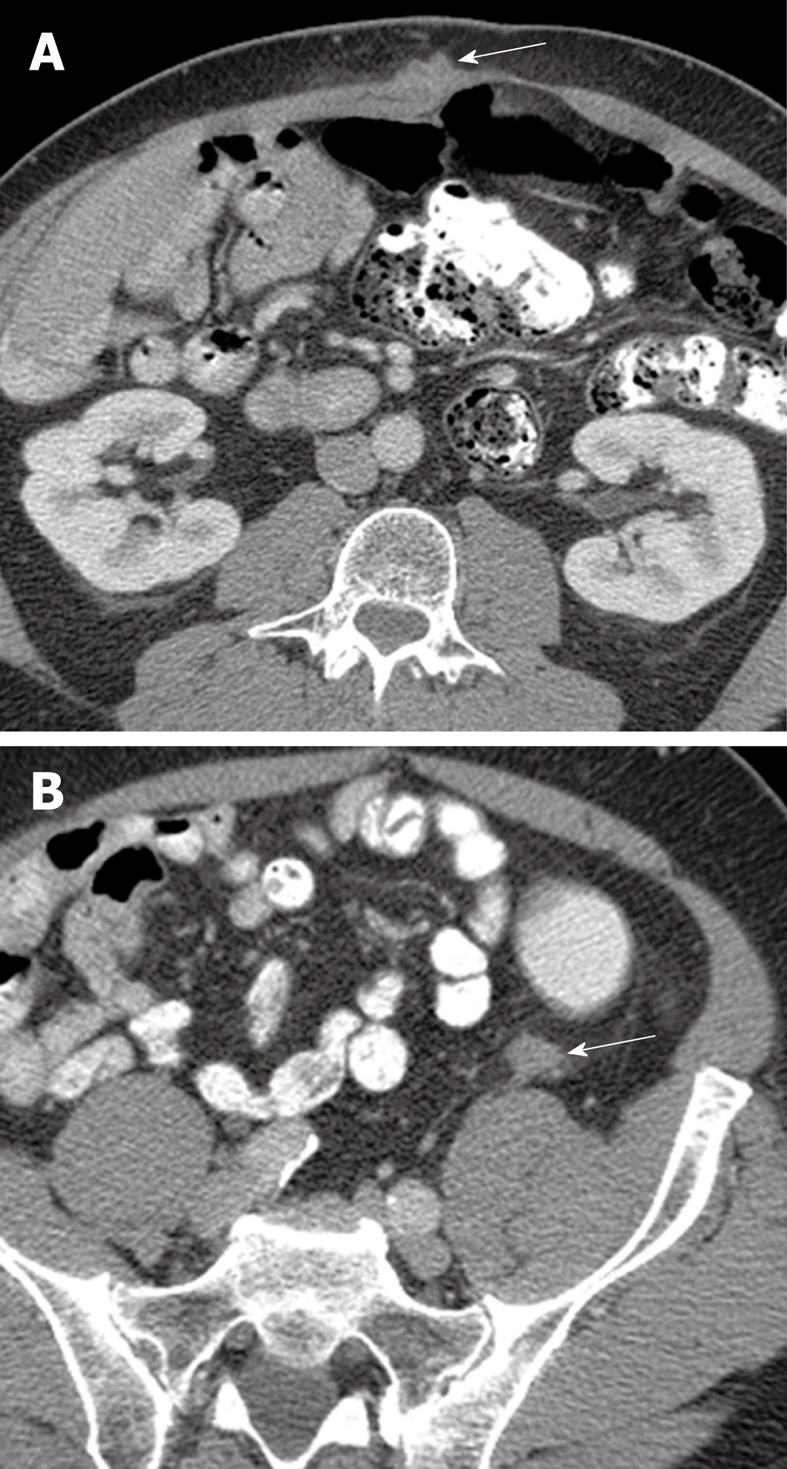
Figure 3.

64-year-old male with metastatic adenocarcinoma of the colon. A: Surveillance axial contrast-enhanced CT image shows a metastatic deposit in the right rectus abdominis muscle (arrow); B: A second metastatic lesion is present in the left paracolic gutter (arrow). The high spatial resolution of CT and the contrast with the adjacent fat allows for easy detection of metastatic disease in these areas.
