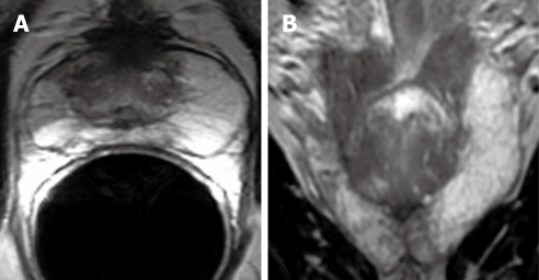
Figure 1.
Magnetic resonance (MR) images demonstrating zonal anatomy of prostate gland. A: Axial T2-weighted (T2W) MR image depicts the central gland and peripheral zone (PZ). Central gland is hypointense compared to hyperintense PZ; B. Coronal T2W MR image shows hyperintense PZ and hypointense central gland.

