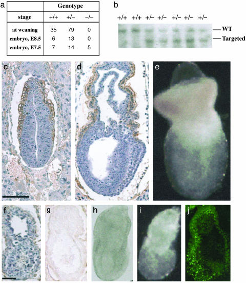
Fig. 2.
Disruption of beclin 1 causes early embryonic lethality. (a) Genotype distribution of offspring and embryos from beclin 1+/- intercrosses examined by Southern blotting or PCR. (b) Southern blot analysis of mouse genotype at age of weaning from a litter born by heterozygous parents. (c and d) Immunohistochemical analysis of beclin 1 expression in wt or heterozygous embryo at E6.5 and E7.5, respectively. Sections were counterstained with hematoxylin. (e) Image of wt whole embryo at E7.5. c–e are at the same magnification with a scale bar of 100 μm. (f and g) Sections from the same null mutant embryo (E7.5) were immunostained with anti-beclin 1 antibody and counterstained with (f) or without (g) hematoxylin. (h) Whole embryo of null mutant (E7.5) viewed under a Zeiss Axiovert confocal microscope with differential interference contrast microscopy. (i) The same embryo as in h viewed under serological microscope. (j) Whole embryo of null mutant stained with acridine orange and viewed with fluorescence under a Zeiss confocal microscope. f–j are at the same magnification with a scale bar of 20 μm.

