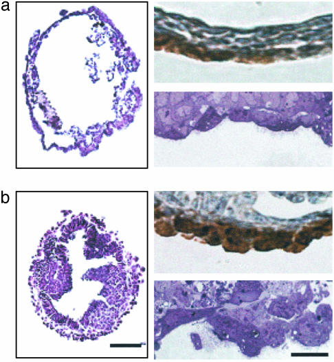
Fig. 3.
Disruption of beclin 1 causes abnormal formation and cell growth in VE of EB. Analysis of EBs (day 14) derived from ES cells of wt (a) and beclin 1-/- (b) is shown. (a and b, Left) H&E staining of paraffin sections. Note the expanded cystic form for wt and the cystic cavitated form for beclin 1-/-. (Scale bar, 100 μm.) (a and b, Right) Sections immunostained with anti-amnionless antibody (Upper) and toluidene blue-stained, 1-μm sections (Lower). (Scale bar, 10 μm.)

