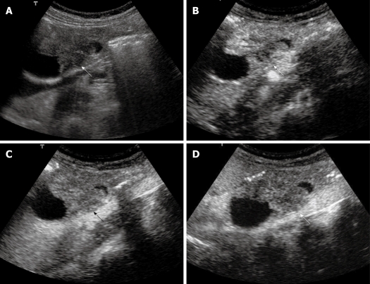
Figure 4.
Extrahepatic cholangiocarcinoma. A: Baseline ultrasound shows an isoechoic mass (arrow) in the lower portion of the common bile duct; B: The lesion (arrow) shows heterogeneous iso-enhancement 13 s after contrast agent injection on CEUS; C: The lesion (arrow) continues to be iso-enhanced 56 s after contrast agent injection; D: The lesion (arrow) becomes hypo-enhanced 107 s after contrast agent injection.

