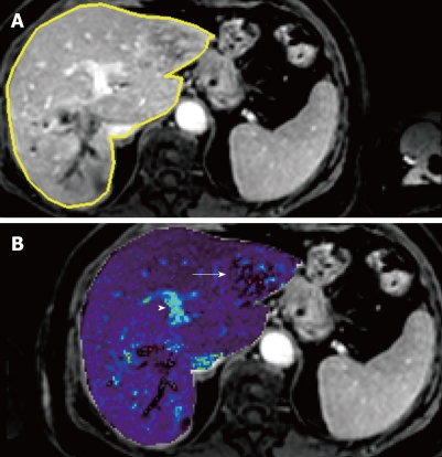
Figure 2.
Liver metastasis in a 68-year-old woman using transverse contrast-enhanced 4D THRIVE. The reference image using 4D THRIVE in the delayed phase (A) is shown with corresponding automatically calculated Kep map (B). The parametric map shows the liver metastasis (white arrow) as a heterogeneous lesion with ring enhancement. The portal vein is indicated by the white arrowhead.

