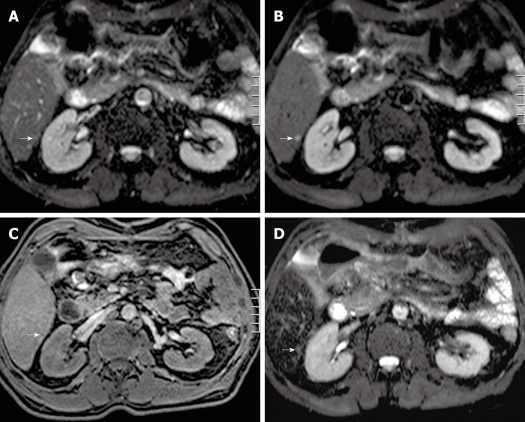
Figure 3.
Transverse single-shot spin-echo echo-planar imaging (SS SE-EPI), fat-suppressed T1w 3D GE and SPIO-enhanced T2w TSE (short TE with fat suppression) image in a 62-year-old man. A: A transverse SS SE-EPI image using b = 0 s/mm2 in a 62-year-old man barely detecting any visible lesion in the area indicated by the white arrow; B: A transverse SS SE-EPI image using b = 10 s/mm2 in the 62-year-old man clearly detecting the liver metastasis (white arrow); C: A transverse fat-suppressed T1w 3D GE image in the portal-venous phase after intravenous injection of SPIO in the 62-year-old man barely detecting the liver metastasis (white arrow); D: A transverse SPIO-enhanced T2w TSE (short TE with fat suppression) image in the 62-year-old man barely detecting the liver metastasis (white arrow). SS SE-EPI: Single-shot spin-echo echo-planar imaging; GE: Gradient echo; T1w: T1-weighted; SPIO: Superparamagnetic iron oxide; TE: Echo time.

