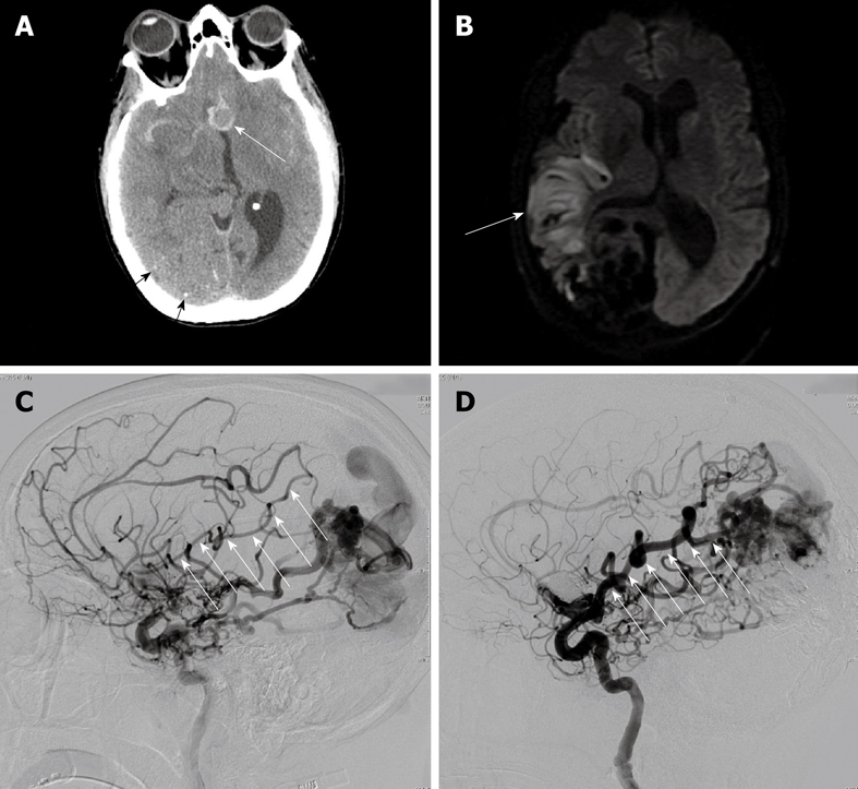
Figure 1.

A 47-year-old woman with ruptured anterior communicating artery aneurysm associated with a large arteriovenous malformation. A: Computed tomography brain scan shows the 12 mm anterior communicating artery aneurysm in relief surrounded by high-density subarachnoid hemorrhage (white arrow). Also note small stippled calcifications in the large parietal-occipital arteriovenous malformation (black arrows); B: Diffusion-weighted magnetic resonance imaging shows area of restricted diffusion in the posterior right frontal lobe (white arrow) corresponding with the patient’s left-sided weakness and hemi-neglect; C: Catheter arteriography of the right internal carotid artery in the lateral projection at the time of initial presentation approximately 1 wk following subarachnoid hemorrhage shows significant narrowing in the angular and Rolandic branches of the right middle cerebral artery, both of which supply the arteriovenous malformation (white arrows); D: Catheter arteriography of the right internal carotid artery in the lateral projection 4 mo after initial presentation and following complete neurological recovery shows resolution of vasospasm in the angular and parietal branches of the right middle cerebral artery supplying the arteriovenous malformation (white arrows).
