Fig. 1.
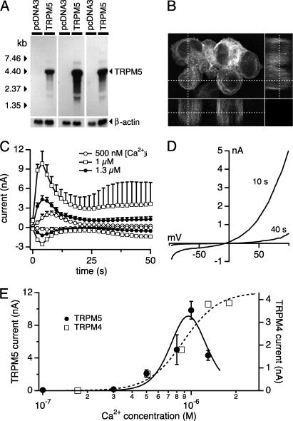
TRPM5 is a transmembrane protein and a calcium-activated cation channel. (A) Northern blot analysis of HEK-293 cells stably transfected with pcDNA3-TRPM5 and pcDNA3. (B) Confocal laser microscopic analysis of HEK-293 cells expressing the EGFP-TRPM5 fusion protein showing significant amounts of the protein in the plasma membrane. (C) Average TRPM5 currents at -80 mV or +80 mV in HEK-293 cells perfused with intracellular solutions buffered to the indicated levels of [Ca2+]i (n = 5–20). (D) Typical I/V curve of TRPM5 currents measured 10 s or 40 s after establishment of whole-cell configuration with 500 nM [Ca2+]i. (E) Concentration–response curve of TRPM5 currents (left axis, filled circles; n = 5–20). The fit represents the product of two Boltzmann functions with an apparent EC50 for activation of 840 nM (Hill coefficient, 5). The right axis shows the dose dependence of TRPM4 currents evoked by different concentrations of intracellular [Ca2+]i (data recalculated by webmaxc, see Methods). The fit to these data yields an EC50 of 885 nM (Hill coefficient, 3.6).

