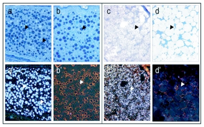
Figure 4.
Yolk bodies in vitellogenic oocytes at recorded at 40 × magnification, (see arrows; bright field: a-d, dark field: a'-d'). Controls (b, b' and d, d') are treated with preimmune sera instead of primary antibody. At the onset of the vitellogenic phase, vitellogenin positive yolk bodies (see arrows) are numerous, small and dispersed through the oocyte (a, a'). Yolk bodies are intensely labeled against vitellogenin, whereas the areas between the yolk bodies show much lower levels of labeling. Mature oocytes, like the one depicted from a stage 4 ovary (mature ovary with at least one egg), are characterized by large vitellogenin positive globules (see arrows) that evolve from yolk body fusion (c, c'). Note absence of labeling in controls (b,b' and d, d').

