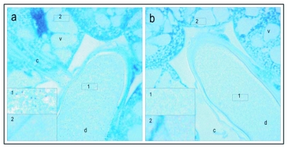
Figure 5.
Verification of presence of mature vitellogenin protein in hypopharyngeal glands of a honeybee worker that performed brood care. Immuno labeling shows that the vacuoles (v), the canal (c) and the common duct (d), are all vitellogenin positive. The antigen is scattered in the gland and in comparison with oocytes there seems to be no aggregation of vitellogenin. In the high resolution micrograph (100 x) (a) labeling is seen as black dots (as exemplified by inserted box 1, higher magnification of stain in the common duct). The antigen is not present in all the vacuoles in a gland (see inserted box 2 in (a), higher magnification of labeling free area in the vacuole). The corresponding preimmune controls (b) show no positive labeling for vitellogenin, neither in the common duct (see inserted box 1 in (b) for higher magnification of the area) or the vacuoles (see inserted box 2 in (b) for higher magnification of the area).

