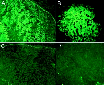
Figure 2.
Wing epidermal cells expressing EGFP after plasmid injection followed by electroporation. A) 3 pulses of 80 V, o.1 ms pulse duration, and 100 ms pulse interval (100X magnification). B) 3 pulses of 80 V, 1 second pulse duration, and 500 ms pulse interval (200X magnification). C) and D) Epidermal cells of wings treated in the same way as in A) and B) but injected with blue colored water.

