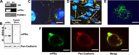
Figure 1.
Presence of PRs and localization of mPRα in granulosa and theca cocultures. A, Detection of PR, mPRα, and PGRMC1 mRNA in isolated granulosa/theca cells using RT-PCR. RT plus and RT minus shown to confirm lack of DNA contamination. B, Western blotting of mPRα (top panels) in two G/T cell plasma membrane preparations using an IgG-purified antibody directed against an N-terminal mPRα peptide (left panel) and blocked with the peptide antigen (right panel). Bottom panels, Pan-cadherin loading control. C–E, Immunocytochemistry of G/T cell cultures using the mPRα antibody and 6-diamidino-2-phenylindole nuclear staining. Scale bar, 2.5 μm (C) and 10 μm (D). Confocal microscopy shows punctate mPRα staining on the cell surface. Scale bar, 5 μm (E). F, mPRα colocalizes with pan-cadherin on G/T cell membranes. Scale bar, 5 μm.

