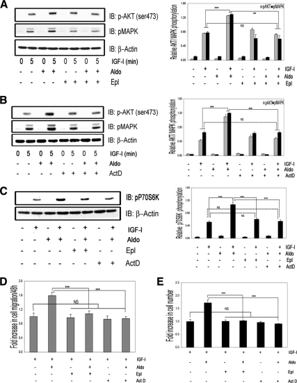
Figure 3.
The enhancing actions of aldosterone on IGF-I signaling and biological function require aldosterone binding to MR and gene transcription. A, VSMCs were incubated with eplerenone (10 μmol/liter) for 1 h, and then 10 nmol/liter of aldosterone for 18 h, or with eplerenone alone, and then IGF-I (50 ng/ml) for 5 min. IB, Immunoblot; Aldo, aldosterone. B, VSMCs were incubated with Act-D (20 nmol/liter) for 1 h followed by aldosterone (10 nmol/liter) for 18 h or Act-D alone, and then IGF-I (50 ng/ml) was added for 5 min. C, VSMCs were incubated with Epl (10 μmol/liter) or Act-D (20 nmol/liter) for 1 h and then 10 nmol/liter of aldosterone for 18 h or with Act-D alone for 18 h and then IGF-I (50 ng/ml) for 5 min. A–C, The levels of phosphorylated Akt, MAPK, and p70S6K were determined by direct immunoblotting and normalized to β-actin. The bar graphs show the mean ± sem (n = 3). ***, P < 0.001; **, P < 0.01. D, Cell migration. Aldosterone (10 nmol/liter), Epl (10 μmol/liter), and Act-D (20 nmol/liter) were added in combination with IGF-I or IGF-I plus aldosterone. ***, P < 0.001, when the response to aldosterone plus IGF-I is compared with IGF-I plus aldosterone plus Epl or to IGF-I plus aldosterone plus Act-D. NS, Not significant when the response to Epl or Act-D plus IGF-I is compared with the response to IGF-I alone. The results are the mean ± sem. E, Cell proliferation. Aldosterone (10 nmol/liter), Epl (10 μmol/liter), and Act-D (20 nmol/liter) were added in combination with IGF-I. ***, P < 0.001 when aldosterone plus IGF-I is compared with the response of cells to aldosterone plus IGF-I plus Epl or to IGF-I plus aldosterone plus Act-D. NS, Not significant when Epl or Act-D plus IGF-I is compared with the response of cells to IGF-I alone.

