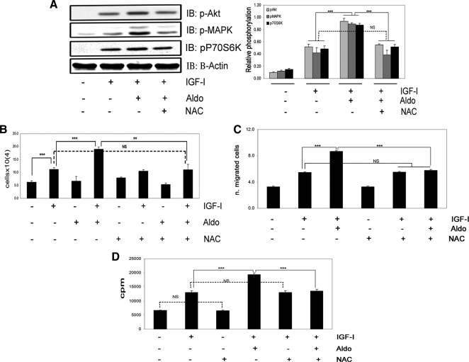
Figure 8.
The enhancing action of aldosterone on IGF-I-mediated signaling and biological function is completely inhibited by NAC. A, VSMCs were incubated for 60 min with or without NAC (2 mmol/liter) and aldosterone (10 nmol/liter) for 18 h before IGF-I (50 ng/ml) was added for 5 min. Levels of Akt, MAPK, and p70S6K phosphorylation were assessed by immunoblotting. The blots were stripped and reprobed with anti-β-actin. The results from three similar experiments are expressed as the ratio between the phosphorylated protein and β-actin. IB, Immunoblot; Aldo, aldosterone. B, Cell proliferation. Cells were treated with or without NAC (2 mmol/liter), aldosterone (10 nmol/liter), and/or IGF-I (50 ng/ml) for 48 h, as indicated. Each data point represents the mean ± sem number of cells (×104) of three replicate measurements from three independent experiments. C, Cell migration. NAC (2 mmol/liter), aldosterone (10 nmol/liter), and/or IGF-I (50 ng/ml) were added for 48 h, as indicated. ***, P < 0.001 when aldosterone plus IGF-I is compared with the response of cells to IGF-I alone or IGF-I plus aldosterone compared with IGF-I plus aldosterone plus NAC. NS, Not significant when aldosterone and NAC plus IGF-I is compared with the response of cells to IGF-I or NAC alone. D, Protein synthesis. VSMCs were incubated for 18 h with or without NAC (2 mmol/liter), aldosterone (10 nmol/liter), and/or IGF-I (50 ng/ml), as indicated. Each data point represents the mean ± sd cpm of two replicate measurements from three independent experiments. ***, P < 0.001. NS, Not significant.

