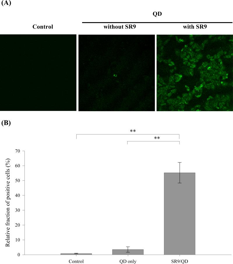
Fig. 2.
Confocal microscopy of cellular uptake of SR9/QD complexes. (A) Confocal microscopic images of cells treated with QD or SR9/QD complexes. Human lung carcinoma A549 cells treated with mock as a control, 100 nM of QD (Evident Technologies, Troy, NY), and SR9/QD complexes at a molecular ratio of 60:1 (6 μM of SR9 peptide premixed with 100 nM of QD) for 1 hour were shown on the left, middle, and right, respectively. All images were recorded by the TCS SL confocal microscope system (Leica, Wetzlar, Germany) and shown at a magnification of 200X. (B) Efficiency of cellular uptake of QD or SR9/QD complexes. Cells were treated with different conditions as described above and analyzed by the Cytomics FC500 flow cytometer (Beckman Coulter, Fullerton, CA). Significant differences were shown for P < 0.01 (**).

