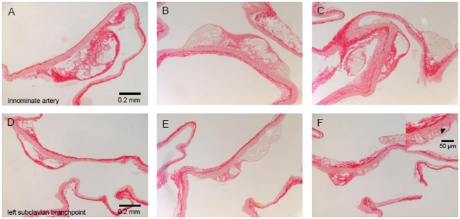
Figure 1. Photomicrographs of Sirius red-stained lesions within two major vessels branching from the aortic arch with a difference in onset of atherogenic development.
The (A, B, C) innominate- (early start) and (D, E, F) left subclavian artery (later start) from apoE−/− mice on (A, D) a Western-type atherogenic diet alone, or in combination with (B, E) inactive- or (C, F) active hyaluronidase infusion, (inset F) Higher magnification of Sirius red-stained lesion within the left subclavian artery from apoE−/− mice on a combined Western-type atherogenic diet and active hyaluronidase infusion. Arrow head indicates absence of long stretches of the intimal layer underneath a plaque. Bar = 0.2 mm, bar inset = 50 µm.

