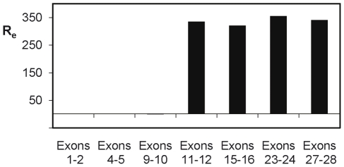
Figure 3. RT-Q-PCR analysis of the expression along the EGFR gene in tumour 26.
For each pair, the forward and reverse primers were localised in consecutive exons in order to prevent amplification of DNA traces present in the mRNA preparations. The expression ratio (Re) was calculated by dividing the normalised expression measured in the tumour by the mean of the normalised expressions measured in the reference set of tumours. Exons 1 to 10 were not over-expressed, indicating that only the truncated form of the gene coded by amplicon 3 was expressed, whereas the large forms present in amplicons 1 and 2 were not expressed (see Figure 1).

