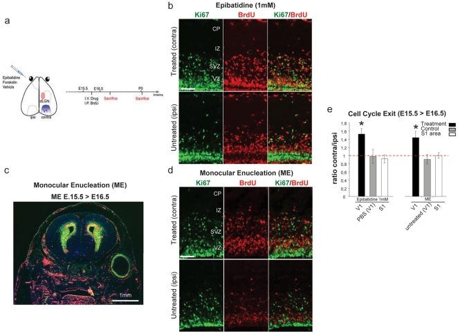
Figure 1. Inhibition of fetal retinal spontaneous activity and Monocular Enucleation (ME) increases corticogenesis.
(a) Schematic representation of the intraocular pharmacological injection to evaluate the effect of the treatment on the contralateral (treated) compared to ipsi-lateral (untreated) developing visual (V1) or somatosensory cortex (S1). The intravitreal (I.V.) injection of epibatidine or monocular enucleation (ME) were performed at E15.5 (supplementary video S1, Figure 1) and BrdU was intraperitoneally (I.P.) administered 4 h later. (b and d) Representative images of Ki67 (green, left panels), BrdU (red, middle panels) and merged labelling (right panels) in contra-lateral (top) and ipsi-lateral (bottom) cortices after epibatidine treatment (b) or ME (d) of E16.5 embryos. Cells withdrawn from the cell cycle are BrdU+/Ki67−. Cells re-entering the cell cycle are BrdU+/Ki67+. (c) Representative image of Ki67 (green) BrdU (red) staining of an E16.5 embryonic head monocularly enucleated at E15.5. (e) Quantification of cell cycle exit rate, reported as the ratio between contra- and ipsi-lateral cortices, in E16.5 embryos upon administration of epibatidine or ME. Epibatidine treatment or ME (epibatidine n = 6, PBS n = 4 p = 0.0004; ME n = 4, untreated n = 3 p = 0.01, Student's t-test, two samples equal variants) increased neurogenesis. None of the treatments resulted in changes of neurogenesis in somatosensory areas (S1, epibatidine on V1 vs S1 n = 4 p = 0.0033; ME on V1 vs S1 n = 4 p = 0.01, Student's t-test, two samples equal variants). Abbreviations: CP cortical plate, IZ intermediate zone, SVZ Sub-ventricular zone, VZ ventricular zone.

