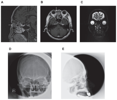
Figure 1.
Preoperative MRI and dacryocysteographic findings in a 77-year-old patient. Both magnetic resonance imaging examinations of T1 images (A, B) and T2 image (C) show a low or isointensity mass in the right lacrimal sac without invasion of surrounding tissues, which is consistent with histopathologic examination; the adjacent epithelium was not invaded by lymphoma cells. Dacryocysteography shows an obstruction of the right nasolacrimal duct (D, E).

