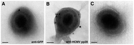
Figure 3. Immuno EM localisation of Rab27a on the viral envelope of isolated HCMV viral particles.
Isolated viral particles from BJ1 YFP-Rab27a cells HCMV infected for 5 days were permeabilised with saponin and labelled with antibodies against YFP (A) and HCMV tegument viral protein pp28 (B), and 10 nm protein-A gold. (C) General morphology of HCMV virions by negative staining with 2% uranyl acetate. When viral particles were partially disrupted, uranyl acetate revealed the nucleocapsids. Scale bar, 50 nm.

