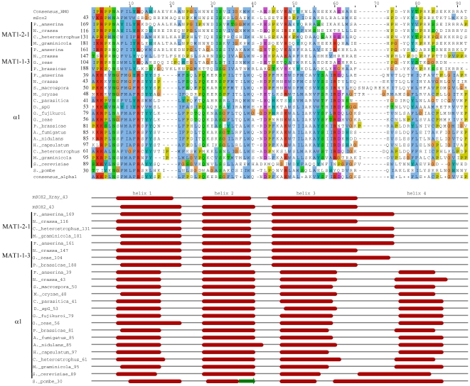
Figure 4. Secondary structure of MATA_HMG and α1 domains from proteins of representative species of Pezizomycotina.
The alignment was obtained with ClustalW2 [63] and coloured according to the Clustal X colour scheme provided by Jalview [64]. This colour scheme is displayed in Table S3. The prediction of secondary structures was performed with Jpred3 [27]. All diplayed helices have a JNETCONF score of at least 7, except for helix 2 from S. pombe which has a JNETCONF score of 0 for all helix 2 positions. The secondary structure presented in the mSOX2_Xray line is from [28] and served to validate accuracy of Jpred3. Full species names and accession numbers are in Table S4.

