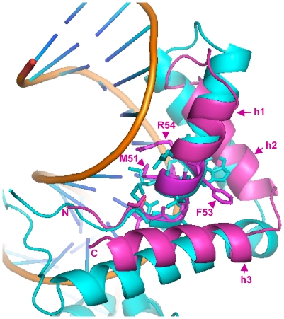
Figure 5. 3D-structure of the α1 domain from MAT1-1-1/mat A-1 of N. crassa.
Schematic ribbon presentation of the superposition of the α1 domain (magenta) onto the structure of the Sox2 HMG domain (cyan) as observed in the tertiary DNA/Sox2/Oct1(POU domain) complex. DNA is represented as gold ribbons (polyphosphate) and blue sticks (bases). Amino acid residues important for DNA recognition and bending are represented as sticks. Residues (methionine M51, phenylalanine F53 and arginine R54) putatively important for function are labelled. Numbering is from the N-terminus methionine. Alpha helices are labelled h1, h2 and h3. Accession number: AAC37478, 3D structure established from residue 44 to 97.

