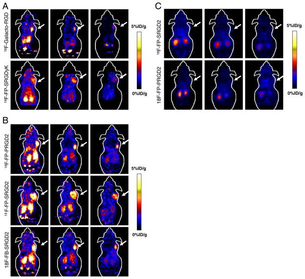
Fig. 3.
Small-animal PET images of U87MG tumor-bearing mice. a Decay-corrected whole-body coronal images at 20 min, 1, and 2 h after injection of about 3.7 MBq of 18F-galacto-RGD and 18F-FP-SRGDyK. b Decay-corrected whole-body coronal images at 20 min, 1, and 2 h after injection of about 3.7 MBq of 18F-FP-SRGD2, 18F-FP-PRGD2, and 18F-FB-SRGD2. c Decay-corrected whole-body coronal images at 1 h after injection of about 3.7 MBq of 18F-FP-SRGD2 and 18F-FP-PRGD2 with coinjection of 10 mg c(RGDyK) per kilogram of mouse body weight

