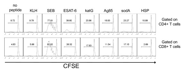
Figure 6.
Sarcoidosis BAL T cells proliferate in response to multiple mycobacterial antigens. Sarcoidosis BAL cells were CFSE-labeled and activated with neoantigen (KLH), ESAT-6, katG, Ag85A, sodA, and HSP. Day 4 post-activation, antigen-specific proliferation of CD4+ and CD8+ T cells was assessed by gating on CD3+CD4+ T cells or CD3+CD8+ T cells and analyzing the CFSE expression of each subset by flow cytometry. Percent proliferation is indicated by bracket above peaks. Similar results were found in the three other tested subjects.

