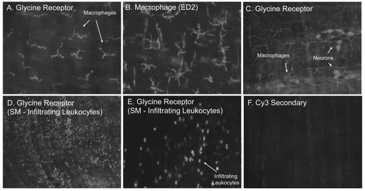
Figure 1.
Panels A-F. Fluorescence micrographs of rat jejunal whole-mounts immunohistochemically stained with a specific antibody against the rat glycine receptor with the secondary conjugated to Cy3. Panel A.) Muscularis whole-mounts from control rats expressed a highly fluorescent immunoreactive signal to the glycine receptor on stellate shaped cells. Panel B.) Using macrophage specific ED2 immunohistochemistry, these cells were identified as macrophages. Panel C.) In addition to macrophages, enteric neuronal cells presented with glycine receptor-like immunoreactivity. Panels D and E.) Numerous glycine receptor positive infiltrating leukocytes were observed 24 hours after intestinal manipulation. Panel F.) No specific immunoreactivity was visualized using a control Cy3 staining without primary antibody incubation. (original magnification: A, C, E and F=200X; B=250X; D=100X).

