
Figure 1. Clinical manifestation of corgi muscular dystrophy.
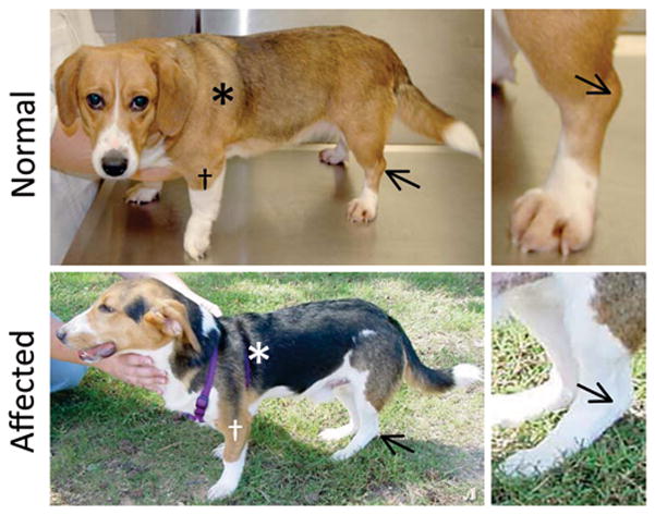
Representative photographs of 10-m-old corgi in standing position. Top, Rachel, a normal corgi. Bottom, Jerry, a dystrophic corgi. Asterisk indicates proximal limb muscle; Cross indicated distal limb muscle; Arrow indicates the tibial-tarsal joint. The affected dog Jerry shows limb atrophy and his tibial-tarsal joints are dropped to the ground.