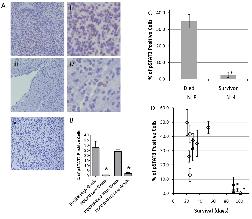
Fig. 2.
(A) Representative light microscopy images showing immunohistochemical staining for p-STAT3 in a high-grade glioma at (i) low (100X) and (ii) high (400X) magnification, and in a low-grade tumor at (iii) low (100X) and (iv) high (400X) magnification, and (v) rabbit IgG isotype control (100X). (B) In the PDGF-B alone, and PDGF-B + Bcl-2 mice that developed gliomas, p-STAT3 levels were significantly higher in high-grade tumors than in low-grade tumors (* P < 0.05). (C) In PDGF-B + Bcl-2 mice with high-grade gliomas, animals that survived past 90 days had significantly lower tumor p-STAT3 expression than animals that succumbed to intracranial tumors before 90 days (** P < 0.0001). (D) Mean p-STAT3 expression in individual mice in the PDGF-B + Bcl2 group bearing high-grade tumors plotted against overall survival time showing long-term survival of all animals with p-STAT3 expression that was < 10%. (* = animals sacrificed after surviving past 90 days. Error bars represent standard error of the mean.)

