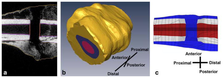
Fig. 1.
a Cortical bone, medullary tissue, and callus are labeled on μCT cans of a sample rat fracture callus. The orientation of the callus is the same as in part c. b 3-D surfaces of the cortical bone, medullary canal, and fracture callus are extrapolated from the labeled images. c The finite element mesh is then contoured to conform to the tissues’ surfaces and scaled. Pictured is a cross section of the finite element model of the fracture callus prior to bending stimulation. Cortical bone is light blue, bone marrow is dark blue, and callus tissue is red

