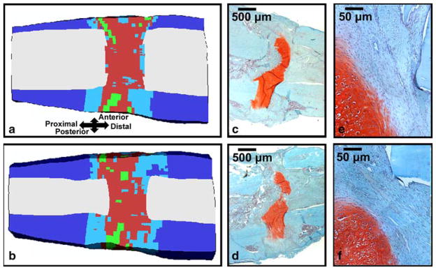
Fig. 4.
a Mid-sagittal and b para-sagittal cross sections of the predicted tissue distribution after four weeks of bending stimulation; the color key is the same as in Fig. 4. c Mid-sagittal and d para-sagittal histology slides (20x magnification) from a specimen from Cullinane et al. (2003) that underwent four weeks of bending stimulation. Regions of (e) and (f) that are adjacent to a cortical fragment and that contain fibrous connective tissue are shown in e and f, respectively (200x magnification). The tissue has been stained with Safranin-O (coloring cartilage red) and Fast Green (coloring bone blue-green)

