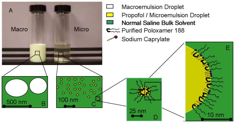
Figure 1.

Photograph and cartoons of a macroemulsion and microemulsion of propofol. Panel A. Shown in the photograph is an opaque, white macroemulsion (Macro) and a clear, colorless microemulsion (Micro) of propofol. Panels B and C: cartoon demonstrating the relative particle sizes of the macroemulsion (Panel B) and microemulsion (Panel C). Panel D. Cartoon of a microemulsion in cross-section. Panel E: Magnified view of cross section of a microemulsion particle demonstrating propofol surrounded by a corona of purified poloxamer 188 and sodium caprylate surfactant in a bulk media of normal saline. Calibration bars apply only to their respective panels. Color legend is applies to all panels.
