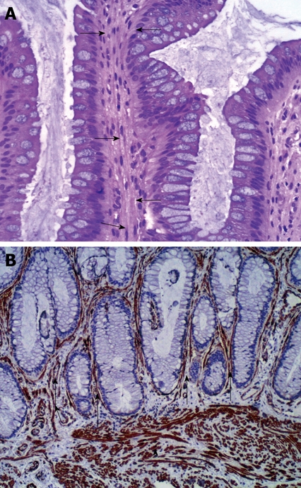
Figure 2.

Histopathological examination. A: The rectal mucosa showing smooth muscle fibers proliferation perpendicular to the muscularis mucosa and extending between the glands (arrows) (HE stain × 40); B: Smooth muscle proliferation in the muscularis mucosa (arrow head as internal control)) and extending in between the mucosal glands (arrows) (Immunohistochemistry, smooth muscle actin, × 100).
