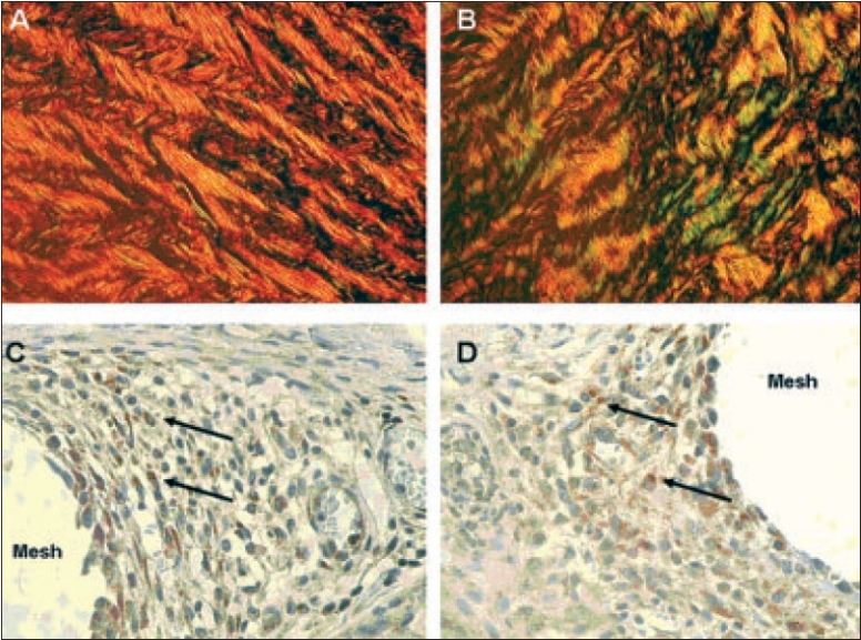
Figure 3.

Cross polarization microscopical (CPM) and immunohistochemical features of human fascial tissue according to Junqueira.[12] CPM of Sirius red-stained section of normal fascia with a collagen type I/III ratio of 14 - (A); and specimen of recurrent incisional hernia fascia with a collagen type I/III ratio of 3.6 - (B). For the detection of MMP-2, we used rabbit polyclonal, 1:1000, from Biomol (Hamburg, Germany) as primary antibody; and goat anti-rabbit, 1:500, from Dako (Glostrup, Denmark) as secondary antibody. Positive cytoplasmatic expression of MMP-2 in granuloma adjacent to mesh filaments - (C, D) (positive stained cells marked with black arrows). (Magnification 400× in images IIIA - IIID).
