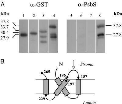
Fig. 1.
Characterization of anti-PsbS serum. (A) Immunoblotting analysis of total extracts of E. coli cells expressing GST alone or GST-PsbS fusion proteins, detected by commercial anti-GST polyclonal antibodies (Left) or anti-PsbS serum produced against recombinant His-tagged PsbS (Right). Shown are GST protein (lanes 1 and 5), GST-G229-E265 fusion (lanes 2 and 6), GST-F197-E265 fusion (lanes 3 and 7), and GST-V157-E265 fusion (lanes 4 and 8). Molecular masses are calculated from amino acid sequences, as deduced from recombinant genes. Two bands detected by anti-GST antibodies under GST-F197-E265 fusion polypeptide (lane 3) are possible products of proteolytic digestion. See text for identification of bands in lanes 4 and 8. (B) Proposed topology of PsbS polypeptide in thylakoid membrane. Number of amino acids defining boundaries of peptides expressed in fusion with GST are shown. Arrow indicates the amino acid loop containing the mapped epitope.

