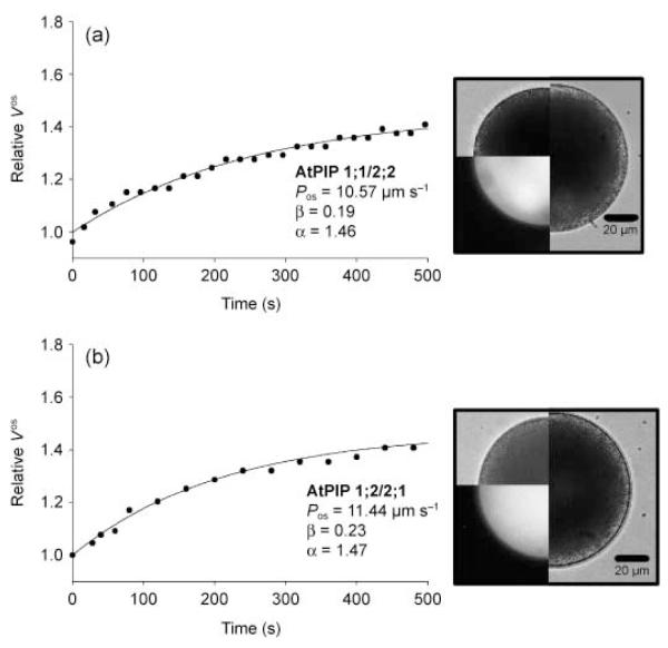
Fig. 5.
Swelling curves of protoplasts obtained from lily (Lilium longiflorum) pollen grains co-transformed with Arabidopsis thaliana plasma membrane intrinsic protein 1;1 (AtPIP1;1)/AtPIP2;2 (a) and AtPIP1;2/AtPIP2;1 (b). The images show the protoplast at t = 0 min as bright field images (upper left quarter) and as fluorescence images (lower left quarter). The right half of the image shows the protoplast at the end of the swelling curve (tfinal). Bars, 20 μm.

