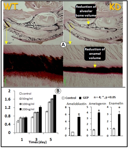
Fig 5.
GEP KD mice display a reduction in enamel mineralization. A) Von Kossa staining showed a reduction of mineralization in the GEP knockdown mice (KD, right panel) compared to the age matched wild type (WT, left panel). B) MTT assay data showed an increase in cell proliferation in the ameloblast cell line with all 3 concentrations of recombinant GEP protein, although there was a significant difference only at the concentration of 200 ng. C) Recombinant GEP protein increased levels of cell markers for ameloblast cell differentiation: ameloblastin, amelogenin, and enamelin. These values were normalized by GAPDH. (Data are mean ± SEM, n=4, p <0.05).

