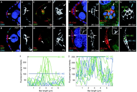
Figure 3.
msn regulates DE-Cad levels in border cells. At stage 8 (S8), (A) WT and (B) msn− border cells localize Dlg (red) at the front, facing the germ line. (C,D) At S9, Dlg is found at high levels at PC–BC and BC–BC interfaces and low levels at the BC–NC interface in both wild-type and msn− border cells. (E,F) DE-Cad distribution in S8 egg chambers is uniform around the surface of both (E) wild-type and (F) msn− border cells. (G,H) At S10, while in wild-type S10 clusters DE-Cad is found at high levels at the PC–BC and BC–BC boundaries and at low levels at BC–NC boundaries, msn− border cells also accumulate high DE-Cad levels at BC–NC interfaces. Insets in F and H are internal controls for DE-Cad staining in follicle cells. (I,J) Histogram of relative fluorescent intensities of DE-Cad along BC–BC (green) and BC–NC (blue) boundaries, as indicated in G′ and H′. Coloured bars indicate the maximum DE-Cad fluorescence intensity found at BC–BC and BC–NC boundaries. Arrows, BC–NC contacts; arrowheads, basal side of border cells. Primed letters are single channels of corresponding merged panels. BC, border cell; DE-Cad, DE-cadherin; Dlg, Discs large; FC, follicle cell; GFP, green fluorescent protein; msn, misshapen; NC, nurse cell; PC, polar cell; WT, wild type.

