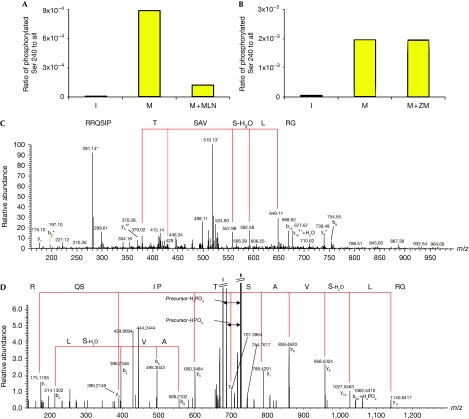
Figure 4.
NUSAP mitotic phosphorylation at Ser 240 correlates with Aurora A activity. Protein samples of FLAG–NUSAP immunoprecipitated from I, M and M+MLN or with M+ZM were analysed using LC-MS/MS, focusing on the predicted phosphorylated residue Ser 240. The histograms (A, B) show the calculated ratios based on peptides carrying the phosphorylated Ser 240 compared with all matched peptides containing this residue. (C, D) MS/MS spectra of two peptides matched the triply charged phosphorylated peptide GRLSphosVASTPISQRR and the doubly charged phosphorylated peptide GRLSphosVASTPISQR, respectively. In both spectra, y-ion series are marked with red lines indicating matched amino-acid sequences (C: doubly charged y-ion series and D: singly charged y-ion series) together with some b ions. The conversion of phosphorylated Ser 240 into dehydroalanine is marked in the spectra. I, interphase cells; LC-MS/MS, liquid chromatography-mass spectrometry; M, mitotic cells arrested with monastrol; M+MLN, mitotic cells co-treated with monastrol and MLN8237; M+ZM, monastrol and ZM447439.

