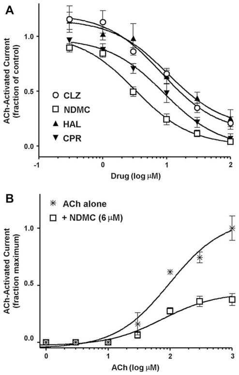
Fig. 2.
(A) Inhibition of acetylcholine (ACh, 60 μM)-stimulated electrophysiological (net charge) responses of Xenopus laevis oocytes expressing rat α7 nicotinic receptors by the antipsychotics clozapine (CLZ) (○), the clozapine metabolite N-desmethylclozapine (NDMC) (□), haloperidol (HAL) (▲) and chlorpromazine (CPR) (▼). Oocytes were voltage-clamped at −60 mV, and data were collected 50 Hz and filtered at 20 Hz. Acetylcholine and drugs were applied (12 s) in the perfusion stream of Ringer’s solution used to bathe (22 °C) the cells. Applications were with ACh alone, then with combination of ACh and antipsychotic, followed by ACh alone for a recovery control. Data are normalized to ACh-alone responses (Means ± S.E.M. for responses of at least 4 oocytes). Curves were generated using nonlinear regression (individual data were best fitted to a one-site model). (B) Insurmountable effect of N-desmethylclozapine (6 μM) on concentration–response relationship of ACh in evoking net charge responses in X. laevis oocytes expressing rat α7 nicotinic receptors. The experimental protocol and data analysis are the same as described for Fig. 2(A) above except that cells were pre-equilibrated with 6 μM N-desmethylclozapine alone for 10 s before the co-application of 6 μM N-desmethylclozapine and varying concentrations of ACh. Data are normalized to ACh-alone maximal response (Means + S.E.M. for responses of at least 4 oocytes).

