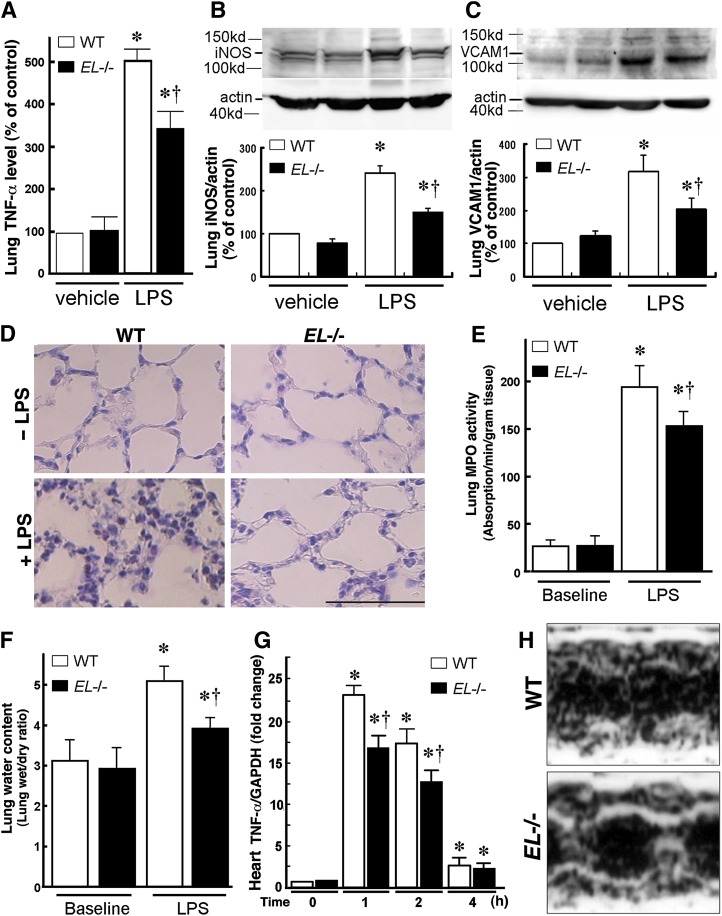
Fig. 5.
EL deficiency attenuates LPS-induced lung inflammation and heart failure. WT and EL−/− mice were injected with LPS (25 mg/kg). After 24 h, the lungs and heart were excised for expression analyses. A: Real-time PCR revealed that the LPS-induced TNF-α mRNA expression in the lung was attenuated in EL−/− mice compared with WT mice. Western blotting revealed that iNOS (B) and VCAM-1 (C) inductions in the lung were attenuated in EL−/− compared with WT mice. Values are expressed as percent of control (WT mice at baseline). D: Lung histology before and 24 h following the LPS injection. Note marked infiltration of polymorphonuclear leukocytes and macrophages in the interstitial spaces and swelling of the alveolar walls, and the LPS-induced damage was attenuated in EL−/− mice. The bar indicates 50 μm. Lung myeloperoxidase (MPO) activity (E) and lung wet/dry ratio (F) were significantly lower in EL−/− mice than in WT mice in response to the LPS administration. G: The LPS-induced TNF-α expression in the heart was attenuated in EL−/− mice compared with WT mice (n = 5 in each group). Bars indicate mean ± SEM. * p < 0.05 versus baseline, † p < 0.05 versus corresponding WT mice (n = 6). H: Representative images of cardiac echocardiography of WT and EL−/− mice after the LPS challenge.

